The SEM electron channelling (EC) photomicrographs shown below provide
an alternative illustration to conventional polarised light and cathodoluminescence (CL)
microscopy of the classic cataclasite fault rock evolution in Cambrian Pipe Rock Quartzite (Skiag Bridge back thrust fault)
via grain size reduction due to fracturing to produce a fine grain size fault rock. In
particular, EC orientation contrast highlights changes in crystal orientation and hence
shows up the intracrystalline (low temperature) plasticity component inherent in
cataclasis. A related image, known as electron channelling patterns (ECP), due to electron
diffraction, not only defines the precise crystallographic orientation but also a
qualitative indication of the amount of cold work hardening present in terms of pattern
quality deterioration.
The photomicrographs are viewed towards NE and were cut normal to fault
plane and parallel to movement direction; where no scale is shown the field of view is
2mm.
For further details, see: Lloyd and Knipe. 1991, Journal of Structural Geology; Knipe
and Lloyd, 1994, Pure and Applied Geophysics 142, 229-254; Lloyd 1987, Mineralogical
Magazine 51, 3-19.
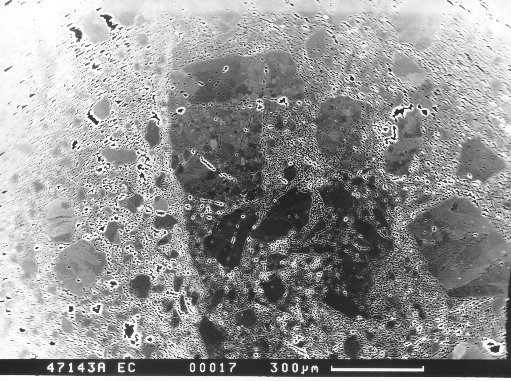
Progressive development of cataclasite as
imaged via SEM electron channelling (EC) orientation contrast The microstructural images
of the cataclastic fault rock developemnet in quartzite in affected by the Skiag Bridge
backthrust fault illustrated on this page were taken using SEM/EC/OC and reflect therefore
changes in crystallographic orientation (Lloyd et al. 1987). Thus, rather than the
fracture and diffusive mass transport (DMT) dominated microstructures as revealed by both
optical and SEM CL, here we see the relationship between intracrystalline (low
temperature) crystal plasticity, fracture and DMT deformation processes and
microstructures.
|
Click on a thumbnail image to see the full
sized picture. Use "back" on your browser to come back to this page.
|
 |
Original Pipe Rock Quartzite unaffected by faulting. Note some
intragranular deformation due to compaction, including dauphin (i.e. penetration) twins
(i.e. dark-bright contrast variations. |
 |
Classic appearance of dauphine twinning due to deformation caused by
stress concentrations at grain contacts due to intergrain indentation. |
 |
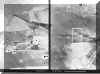
Right. Intragranular LTP 'shear zone' (central grain) due to
indentation by lower grain; note displacement of grain boundary accommodated by the
dextral shear sense. Left. Detail of 'shear zone'; the 'stripes' share
dauphine twin relationship. |
 |
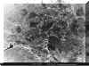
The classic intergranular LTP 'indentation-related' microstructure in
quartz. An inherited and/or initial microstructure (including dauphine twins) in the
central grain has been overprinted by deformation lamellae that originate from intergrain
contacts; several distinct lamellae stes are therfore distinguished. The central grain has
indented the top grain to produce an arcuate array of subgrains; this comminuted 'grain
size' has profound implications for local DMT processes. |
 |
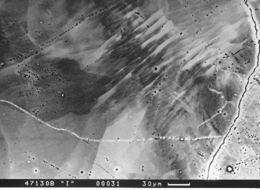
LTP intragranular deformation lamellae caused by indentation of the lower
right grain into the central grain. In detail, the lamellae may be microstructurally
analogous to 'chevron folds' (dauphine twins?) or imbricate thrust stacks. |
 |
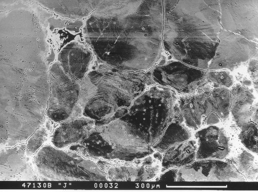
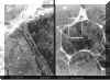
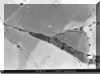
Cataclastic 'seam'; note variations in fragment size and relict quartz
grains in the adjacent wall rock, one of which (lower right) is 'spalling' fragments into
the 'seam'. |
 |
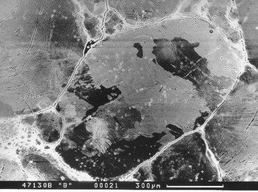
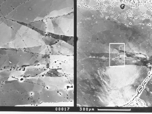
Right. Wall rock grain adjacent to cataclastic seam (top); note
intragranular fractures (cataclasite) that will eventually allow the top of this grain to
'spall' into the main cataclastic seam.
Left. Detail showing intragranular fracture array cutting learlier LTP subgrains
caused by indentation (see opposite image). |
 |
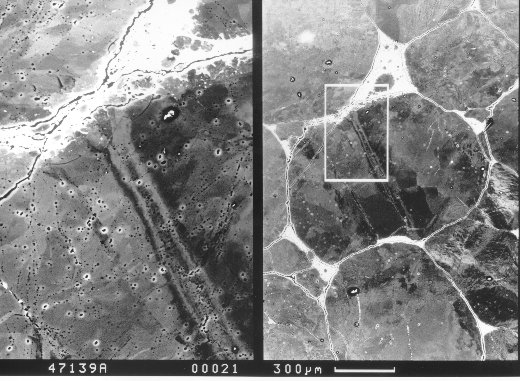
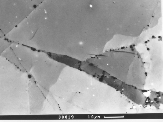
Detail of images opposite, showing fracture bounded 'tilt blocks' that
overprint LTP subgrain boundaries caused by earlier indentation. The fractures have
the appearance of a propagating crack-tip (from right-to-left), indicating dextral offset
of the main subgrain boundary. The LTP therfore may represent crack-tip plasticity
associated with an earlier stage of propagation. |
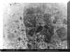 |
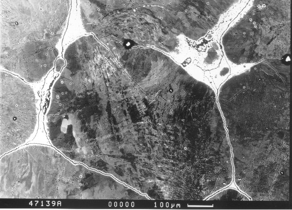
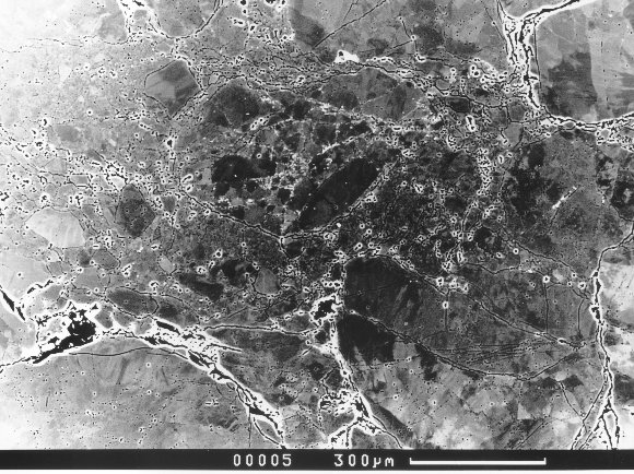
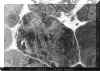
Crystal plastic microstructure of the main cataclastic fault rock. Note
contrast variations due to differences in fragment sizes that impart a 'foliated'
appearance |
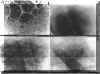 |
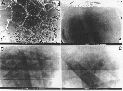
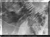
Electron channeling patterns from different grains progressively closer
(lower left, lower right, top right) to a cataclastic seam show progressively poorer
quality due to increased cold work hardening. |