Cathodoluminescence images of cataclastic fault rock evolution
The cathodoluminescence (CL) photomicrographs (both optical microscope and SEM derived)
shown below provide an alternative illustration to conventional polarised light microscopy
of the classic cataclasite fault rock evolution in Cambrian Pipe Rock
Quartzite (Skiag Bridge back thrust fault) via grain size
reduction due to fracturing to produce a fine grain size fault rock. In particular, CL
highlights intragranular microfractures and authigenic quartz precipitation (usually dark
contrast). Note that optical CL images are true colour, but SEM CL images are merely
contrast (signal strength) grey scale variations.
The photomicrographs are viewed towards NE and were cut normal to fault plane and
parallel to movement direction; where no scale is shown the field of view is 2mm.
For other examples see: optical,
SEM, EBSD, TEM,
indentation.
For further details, see: Lloyd and Knipe. 1991, Journal of Structural Geology, and
Knipe and Lloyd, 1994, Pure and Applied Geophysics.
|
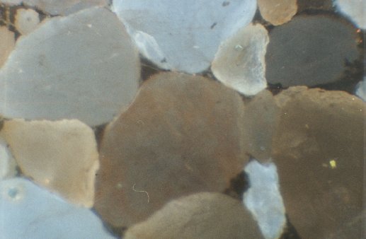
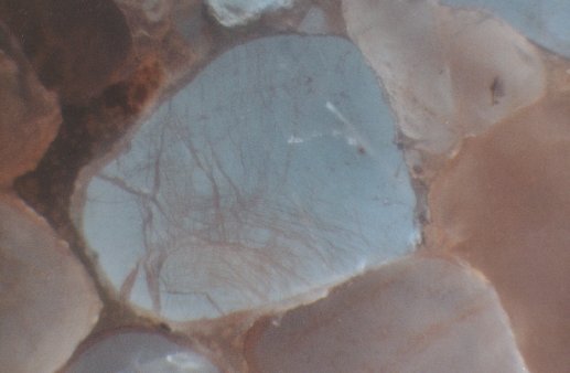
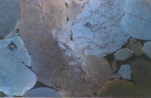
Relatively undeformed quartzite far from the main fault
with a single through-going microcrack. Note: diffuse crack tip termination (symbol left)
and step between grains (arrowed right). The effective crack width (double arrow) if
shearing occurs will be greater than the width observed. |
|
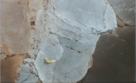
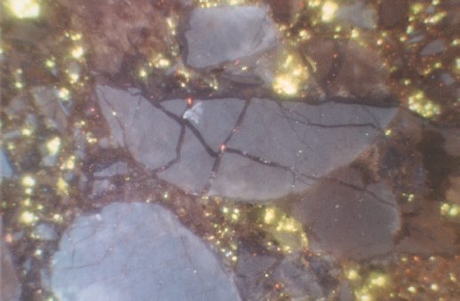
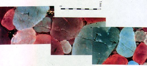
Cataclastic fault rock (right) and a relict fragment of
original quartzite (left) from the main displacement plane. Note: healed (dark)
through-going microcracks in the relict fragment; 'spalling' of grains on the edge of the
fragment into the cataclastic zone, resulting in a weakly foliated (on the basis of
original CL colour index and hence parental grain affinity) cataclasite. |
|
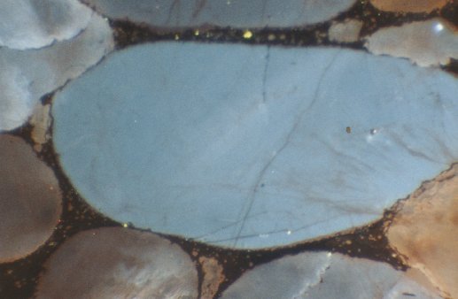
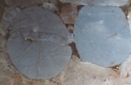
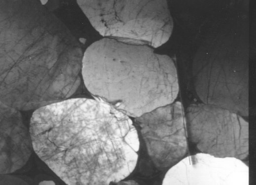
SEM CL image of the wall rock quartzite. Note the
intensity of healed intragranular microcracks and the concentration of deformation at
intergrain contacts due to stress concentration. Same scale as image below. |
|
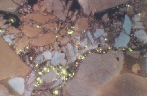
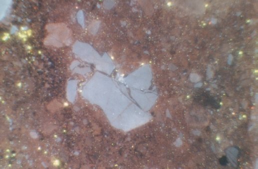
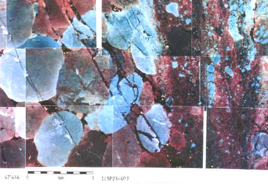
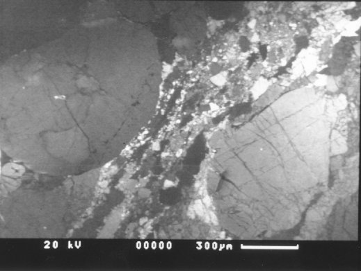
SEM CL image of a narrow cataclastic 'seam' developed
in wall rock quartzite. Note: foliated appearance of the cataclasite due to CL
contrast affinity with the CL contrast of the parental grain; and the 'spalling' of
parental grains on the edge of the seam into the cataclastic fault rock. |